FRCR Part 2B (Radiology) - guidance notes for candidates
LEARN MORE1 Exam structure
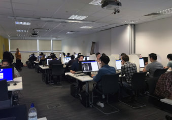
The Final FRCR (Part B) exam comprises three components: a reporting session, a rapid reporting session and an oral exam. All components are examined by an image viewing session delivered on individual workstations.
2 Reporting and rapid reporting components
2a Reporting content
The reporting session comprises six cases, each of which requires a typed report, and it runs for 75 minutes. Each case may comprise multiple modalities including CT, ultrasound, radionuclide and MR scans. Cross-sectional imaging may comprise more than one sequence, which can be scrolled through.
Brief case histories and other relevant clinical data for each case will be displayed, and responses should be presented in a standard format as follows:
- Observations: This section is for recording observations from all the imaging studies available, including relevant positive and negative findings.
- Interpretation: This section is for stating interpretations of the observed findings; for example, describing whether the mass or process observed is benign, malignant or infective rather than neoplastic, giving reasons.
- Main or Principal Diagnosis: This single diagnosis should be based on the interpretations provided above. If a single diagnosis is not possible, then the most likely diagnosis should be stated with a list of other possibilities, in order of likelihood, supplied in the differential diagnosis section below.
- Any Differential Diagnoses: For some cases there will be no differential diagnoses; in others a few may merit inclusion. These should be limited in number and brief, and the report should indicate why these were less likely than the main or principal diagnosis above.
- Any Relevant Further Investigations or Management: This section is for indicating any further appropriate investigations or clinical management. For example, if a patient with a sub-dural collection is diagnosed then urgent referral is needed if there is evidence of brain compression. Similarly, if an abscess or tumour is diagnosed, indicate if drainage or biopsy is appropriate.
The cases vary in complexity and difficulty; some require more time for analysis and reporting than others. Candidates should ensure sufficient time is allocated to report each case adequately. It is not essential to record an answer in every section, candidates will not be penalised for entering their responses in the wrong section, or for entering their responses entirely in one section.
2b Rapid Reporting content
The rapid reporting session comprises 30 cases and it runs for 35 minutes. It requires candidates to identify those cases that show normal appearances and those that show an abnormality. Many cases are similar to those encountered in the reporting of A&E and GP- referred cases; the images are primarily plain radiographs.
Where an abnormality is present, candidates are expected to briefly identify this or give a diagnosis. Each abnormal case shows one significant diagnosable abnormality. Abnormalities in the Rapid Reporting component are not complex and therefore differential diagnoses should not be given. Anatomical variants should be recorded as ‘normal’, and some cases may show minor age-related changes only which should also be recorded as ‘normal’.
Each case is displayed on a single screen, with some cases showing a montage of different views, or a single view only.
2c Equipment and software
The Reporting and Rapid Reporting exam platform allows the provision of image-based exams, and capture of candidate responses, electronically. The platform provides a simple image viewing window and the ability to move through cases. Candidates will record their responses directly via a keyboard and mouse onto the platform.
A demonstration site is available via the College website and enables candidates and trainers to familiarise themselves with the platform in advance of the exam.
During the exam, keystrokes and screen activity are monitored and recorded centrally. If a candidate continues to type after the end of the exam, this information will be recorded, and the College will investigate further to determine whether the candidate should be disqualified. In the unlikely event of computer hardware or software failure during the exam, candidates should alert an invigilator by raising their hand – spare workstations are available if necessary.
3 Oral component
3a Content
The oral exam lasts for 60 minutes in total, during which time the candidate spends 30 minutes with each of two pairs of examiners (and so will be assessed by four radiologists in 15-minute blocks). The oral exam allows for four independent judgments of candidate performance.
A wide range of material of varying complexity will be shown. A higher level of performance will be expected in the interpretation of common and routine exams than will be the case with more complex investigations. Examiners will endeavour to ensure that candidates are shown examples from most of the major clinical radiology sub-specialties and candidates are given the opportunity to demonstrate their powers of observation and deduction. A logical and informed approach to image interpretation, as well as a clear ability to debate the merits, relevance and role of techniques that might assist in further investigation of diagnostic problems, will be expected. Examiners may ask supplementary questions to further assess a candidate’s understanding of the problem.
In reaching a conclusion, candidates should place their diagnoses in order of probability. In some cases, it will be possible to make the correct diagnosis as soon as the signs are elicited.
In others, further views or investigations will be helpful and it is important that candidates clearly state their reasons for wanting these.
Candidates should listen carefully to any information provided and ask for clarification if anything the examiner says is unclear. The amount of discussion that takes place on each case, and the number of cases shown, will vary and is at the discretion of the individual examiner.
3b Equipment and software
The oral component will be delivered to candidates in venues, via remote video conference (MS Teams).
Images are presented digitally on monitors and a practise case will be shown first so that candidates can try scrolling. The examiners will select, display and move through individual images. Exam content will be shared via MS Teams and candidates will be able to take control of the mouse and access functionality to manipulate images. The menu bar for candidate screens will be restricted to zoom, window, pan, scroll, and reset and candidates can use the mouse pointer to indicate areas being discussed.
4 Candidate identification
Candidates are required to bring their candidate timetable to the exam, together with a form of photographic identification. Candidates that are not registered with the General Medical Council must bring their passport. Details on identification documents must match those supplied at application.
The oral examiners identify candidates using their candidate number only. The examiners may introduce themselves by name; however, candidates should not give their name, or any other personal details, in reply.
5 Anonymity of patients
During the exam, information about patients will become available. Candidates are reminded that patients' confidentiality must be respected at all times. Exam cases must not be discussed with anyone other than the examiners.
Patient and hospital identification names and numbers have been removed from the material used in the exam. The examiners advise candidates of any relevant personal details about the oral images under consideration so candidates need not spend time looking for these on the cases shown.
6 Marking and results awarding
6a Reporting and rapid reporting
The platform uses automated marking, which is programmed with acceptable Rapid Reporting answers. The answers will be matched with candidate responses and marks awarded by the system accordingly. Any answers that do not exactly match those within the platform will be reviewed by examiners and awarded an appropriate mark. Final FRCR Part B examiners will review all responses prior to results awarding.
6b Orals
Candidates have the opportunity to gain a maximum of eight marks from each examining pair, and scores are awarded outside of the written exam platform.
6c Overall
Each of the exam’s three components are independently marked, with the three sets of marks considered as a whole to generate a pass or fail - there is no concept of passing one part (e.g. orals) only. Please refer to the scoring system webpage for more information on how results are determined.
7 Feedback for unsuccessful candidates
Candidates undertaking specialty training in the UK or who are designated NHS contributors who are unsuccessful at the exam on two or more occasions will receive written feedback from the Examining Board Chair or Vice-Chairs. This will include a review and summary of performance over the sittings and will also be copied to the TPD and RSA. It is hoped that this will be of assistance when preparing for a further attempt at the exam.
8 Further information
- Queries arising from this webpage should be addressed to the RCR’s Exams Office, either by email to [email protected] or by telephone on 020 7406 5905.
- Queries at the time of the exam should be raised with the invigilators or College staff present.
- Comments, feedback or complaints following the exam should be directed to the Exams Office, either by email to [email protected] or by telephone on 020 7406 5905.
Our exams
Find out more about our FRCR exams in clinical radiology and clinical oncology, and DDMFR exams in dental and maxillofacial radiology.
